Breakthrough in Large Brain Tumor Surgery: Continuous Functional Mapping Applied
Viet Duc Hospital Successfully Implements Electrode Placement Technique Under General Anesthesia to Remove Diffuse Glioma
Viet Duc University Hospital recently treated a patient (43 years old) diagnosed with a large diffuse parenchymal brain glioma (approximately 8cm) in the right hemisphere. The tumor had diffused deep into critical functional areas, including the motor area, language area, and the internal capsule—the region controlling movement on one side of the body.
Medical Challenges and Surgical Standards:
The surgical standard for high-grade malignant gliomas recommends maximal resection of the tumor plus an additional 4-5mm of invasive tissue to minimize recurrence. However, since the patient’s tumor diffused across nearly the entire hemisphere and encroached upon functional areas, the challenge was how to achieve maximal tumor removal while preserving neurological function.
Novel Breakthrough Technique:
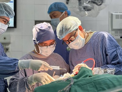
For the first time at Viet Duc University Hospital, the surgical team successfully applied the technique of placing electrodes for continuous functional monitoring while the patient was under general anesthesia. This method replaced the traditional awake craniotomy, which is difficult to apply for tumors of such a large size and long operating duration.
The surgical procedure was strictly implemented:
-
Approach Guidance: Cortical mapping was used to guide access through the “non-functional” area.
-
Continuous Monitoring: Upon reaching the Central Grey Matter (containing the motor pathways), the surgeon used exploratory electrodes to simultaneously suction the tumor and detect functional signals. The electrode system was connected to corresponding muscle groups to monitor motor responses in real-time (right hemisphere surgery monitored the left arm/leg, and vice versa).
-
Precise Warning: When approaching the functional area at about 3-4 mm, the device issued a warning, and at 1mm away, the signal was directly transmitted, allowing the surgeon to stop at the precise moment, thus maximally preserving neurological function while achieving maximal tumor resection.
Outcome:
Thanks to this technique, the team maintained safety even with the tumor encroaching upon nearly the entire hemisphere. Electrical stimulation before wound closure confirmed the patient’s limbs were still moving well. Post-surgery evaluation confirmed the patient had full preservation of motor function, despite the very wide diffusion of the tumor.